Dicke Fellow in Experimental Physics, Princeton University
Fellow, Center for the Physics of Biological Function
Ph.D., University of Southern California
I'm interested in pushing the boundaries of optics to advance both basic science and translational research.
Recent projects focus on inventing and refining microscope technologies to explore how structure and function emerge in complex living systems, from the dynamics of single molecules to computation in the brain.
Favorite papers are highlighted.
By applying two-photon light-sheet microscopy with visual stimuli to larval zebrafish, we map whole-brain activity at cellular resolution and trace the early emergence of "number neurons," revealing that the dynamics of neural networks can encode computation and abstract cognition in the brain.
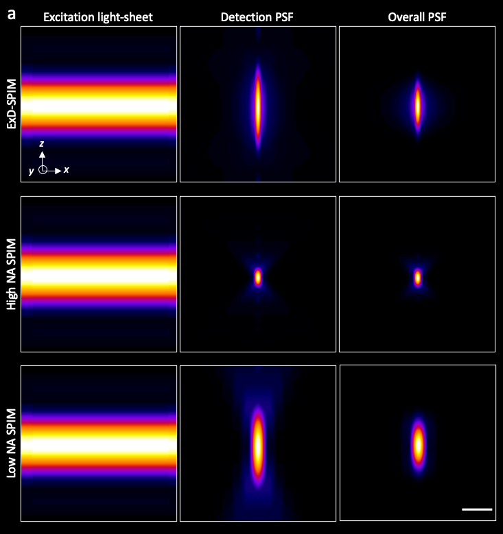
We present a light-sheet approach that maximizes photon collection, and document that this improves the signal-to-noise ratio and volumetric coverage. Our method uses a pupil phase mask to extend the detection depth of field to match the thickness of the illumination light sheet, thereby collecting as useful signal the fluorescence photons that would otherwise be lost or appear as SNR-destroying background.
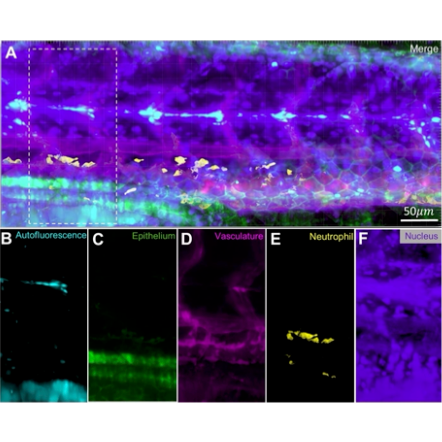
A Fourier transform-based method for snapshot hyperspectral imaging enables photon-efficient and fast fluorescence multiplexing, even in low signal-to-noise conditions.
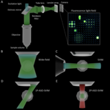
We adapt selective-volume illumination microscopy (SVIM) to a single-objective geometry, using an oblique one-photon illumination path or two-photon excitation to enable high-contrast light-field imaging.
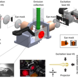
We present the design and construction of an instrument with two independently controlled light-sheet microscope-twins sharing the same multi-laser source, dramatically cutting the cost of the system. We image a variety of specimens, demonstrating instrument versatility and application-specific customization.
This technology is serving researchers at the USC Translational Imaging Center, and has also been adopted by research groups throughout the U.S.
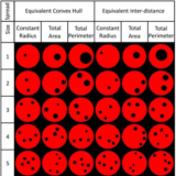
We introduce SVIM, combining the high-contrast associated with light-sheet microscopy and the high-synchronous volumetric acquisition rates of light-field microscopy (limited only by the detector and available signal).
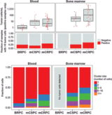
We discuss progress in methods to measure circulating tumor cells, the hematogenous phase of cancer, and we highlight some initial applications, as well as its performance in early-stage diagnosis and treatment monitoring.
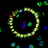
We characterize tumor cells in the fluid phase of blood and bone metastases, revealing new insights into their biophysical properties and clinical potential.
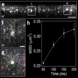
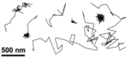
We use split-fluorescent proteins and their activation by complementary synthetic peptides to localize individual molecules and track their diffusion with a precision of 30 nm, directly in live animals.